|
|
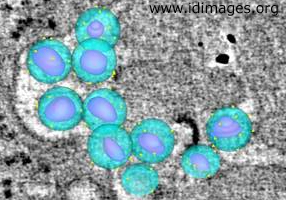
Image from atlas entry: Human immunodeficiency virus (HIV-1 and HIV-2)
Figure 2. Detail of Figure 1, showing modeled HIV-1 virons colorized to show envelope (blue), bullet-shaped core (magenta) and presumed surface spikes (yellow).

- Alternative Image Resolutions
-
- Associated Organisms
- Organ System
- Image Technique
- Microscopy, Electron
- Electron Microscope Tomography
- Related Images
- View other images with a common diagnosis or organism.
- Other Notes
- Image provided by Mark S. Ladinsky and Pamela J. Bjorkman, PhD, of the California Institute of Technology, Pasadena, CA and Douglas Kwon, MD PhD, of the Ragon Institute of MGH, MIT and Harvard and Harvard University, Cambridge, MA.
- URI
- http://www.idimages.org/images/detail/?imageid=1884
|
|