|
|
Image from atlas entry: Human immunodeficiency virus (HIV-1 and HIV-2)
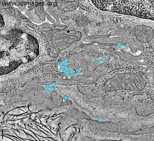
Figure 1. Virions of HIV seen by tomographic electron microscopy in HIV-1 infected BLT mouse colon, with the virion components modeled and highlighted in color.

- Alternative Image Resolutions
-
- Associated Organisms
- Organ System
- Image Technique
- Microscopy, Electron
- Electron Microscope Tomography
- Related Images
- View other images with a common diagnosis or organism.
- Other Notes
- A thin slice from a montaged tomographic reconstruction of a region of a crypt of Lieberkühn in HIV-1 infected BLT mouse colon is shown. The virions have been modeled in position (the envelopes as blue spheres, the bullet-shaped cores in magenta and presumptive surface spikes as tiny yellow spheres) and the tomographic slice superimposed beneath them. This view allows for the virions in the intercellular spaces between cells to be visualized relative to the surrounding tissue components.
Image provided by Mark S. Ladinsky and Pamela J. Bjorkman, PhD, of the California Institute of Technology, Pasadena, CA and Douglas Kwon, MD PhD, of the Ragon Institute of MGH, MIT and Harvard and Harvard University, Cambridge, MA.
- URI
- http://www.idimages.org/images/detail/?imageid=1883
|
|