 |
A man with diffuse large B-cell lymphoma presents with multiple mass like lesions |
- History of Present Illness
- A man in his 70s with intestinal diffuse large B-cell lymphoma receiving chemotherapy presents with fever, chills, and multiple mass-like lesions throughout his body over a 3-month duration. Prior to these symptoms, he had noticed a bruise on his flank with an underlying palpable mass. He had initially attributed this finding to his lymphoma because after his chemotherapy it had decreased in size. However, one day prior to presentation, he bumped himself, leading to purulent drainage from the skin lesion. Throughout this course, he also noticed similar lesions on his mid-scapula and his opposite shoulder, both of which had progressively increased in size despite chemotherapy.
- Past Medical History
- Diffuse large B-cell lymphoma of the bowel, status post R-CHOP, intrathecal chemotherapy, and CAR-T therapy.
- Medications
- Acyclovir, lenalidomide, ondansetron
- Epidemiological History
- The patient lives on a ranch in the desert, located in the Southwestern region of the United States. He has worked with tractors and soil in the past. He owns cattle, dogs, and previously owned horses. He drinks alcohol occasionally and denies ever using tobacco or recreational drugs.
- Physical Examination
The patient appeared well-developed, non-toxic appearing, and in no acute distress. Integumentary exam revealed well-circumscribed, nontender, indurated, erythematous mass-like lesions on his right scapula, and left shoulder. There was also an additional similar appearing lesion in his lower abdomen that had purulent drainage (Figure 1). His chest port site was without tenderness, erythema, or drainage.
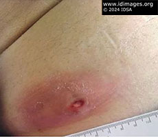 - Figure 1: A well-circumscribed, nontender, indurated, erythematous nodular lesion is seen on the patient’s lower abdomen.

- Studies
On admission, labs were significant for a white blood cell count of 8.39 K/cumm (reference range: normal low 4.00 K/cumm, normal high 10.90 K/cumm), hemoglobin of 10.2 g/dL (reference range: normal low 13.3 g/dL, normal high 17.7 g/dL), platelet count of 219 K/cumm (reference range: normal low 150 K/cumm, normal high 450 K/cumm), sodium of 135 mmol/L (reference range normal low 136 mmol/L, normal high 145 mmol/L), potassium of 3.3 mmol/L (reference range normal low 3.4 mmol/L, normal high 5.2 mmol/L), calcium of 8.3 mg/dL (reference range normal low 8.8 mg/dL, normal high 10.2 mg/dL), albumin of 3.0 g/dL (reference range normal low 3.5 g/dL, normal high 5.2 g/dL), and total protein of 5.6 g/dL (reference range normal low 6.4 g/dL, normal high 8.3 g/dL).
A computed tomography (CT) scan of the abdomen showed a 4.8 x 2.0 cm soft tissue mass in the right lower abdominal subcutaneous fat abutting the skin (Figure 2). In addition, a 2.5 cm soft tissue nodule anterior to the inferior left rectus abdominis muscle and an ill-defined 3.8 x 1.1 cm soft tissue thickening/stranding anterior to the inferior right rectus abdominis muscle were seen. A 2.3 cm soft tissue nodule in the prepubic subcutaneous fat was also identified.
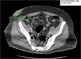 - Figure 2: CT abdomen showed a 4.8 x 2.0 cm soft tissue mass in the right lower abdominal subcutaneous fat abutting the skin.
- Clinical Course Prior to Diagnosis
- Blood cultures were obtained. An incision and drainage of the right lower abdominal soft tissue mass was performed with immediate return of 30 cc of purulent material, which was sent for bacterial, fungal, and acid-fast bacilli culture.
- Diagnostic Procedure(s) and Result(s)
- Wound culture from the patient’s incision and drainage procedure grew Rhodococcus equi (R. equi). One day later, his blood culture from admission also grew Rhodococcus equi as well.
- Treatment and Followup
- After initial cultures grew Rhodococcus equi, the patient was started on azithromycin, vancomycin and imipenem. The blood cultures had initially cleared immediately; however, several days later he had recurrent Rhodococcus equi bacteremia. His chest port was removed. A CT head (with and without contrast) and chest (with contrast), transthoracic echocardiogram (TTE), and transesophageal echocardiogram (TEE) were performed to help identify additional sources of infection. However, they were all unremarkable. Given his persistent bacteremia, the patient underwent aspiration of his right scapular lesion with eventual placement of a drain. After return of the Rhodococcus culture sensitivities, he was discharged on ceftriaxone, azithromycin, and ciprofloxacin for an anticipated two-month course dating from his first negative blood culture. The patient finished his antibiotic course, and on follow up he had significant improvement but incomplete resolution of abscesses. Around 3 months after being discharged, he had repeated incision and drainage of the right lower abdominal quadrant abscess and right upper back abscess, with negative wound cultures and blood cultures.
- Discussion
Risk factors for R. equi infection include immunosuppressive states, including from HIV infection, chemotherapy use, organ transplantation, and diabetes. However, R. equi can notably cause disease in both immunocompromised and immunocompetent hosts (5).
There are different manifestations of R. equi infection. R. equi is a facultative intracellular pathogen that infects macrophages and survives inside lysosomes, and thus can cause chronic infection. R. equi infects a variety of tissues, including the lung, blood, brain, skin, lymph nodes, and bone (1). Most commonly, it causes pneumonia. Complications include development of lung abscess, pleural effusion, empyema; it can also cause complications such as pneumothorax, pericarditis, cardiac tamponade, and mediastinitis (2). Extrapulmonary infections manifest as wound infections, traumatic septic arthritis, and endophthalmitis after ocular injuries. With extrapulmonary infections, the infection usually remains localized at the primary site.
The treatment of R. equi can be difficult (5). R. equi is usually susceptible to vancomycin, macrolides, fluoroquinolones, rifampin, teicoplanin, carbapenems, aminoglycosides, and linezolid. In animal studies, vancomycin, imipenem, and rifampin were the most active antibiotics (3). R. equi is often resistant to penicillins. Monotherapy is generally not recommended because resistance can develop while on therapy. There is some evidence to suggest that combination therapy is more effective than monotherapy, with most experts recommending combination therapy (4). Some experts suggest using a combination of antibiotics with bactericidal and intracellular killing properties for initial reduction of bacterial load (4).
Our patient’s case is a reminder of the importance of obtaining a thorough history. The patient’s demographic history was significant for ranch and horse exposure, which were likely the sources of his infection. Our case also highlights this pathogen’s ability to infect a variety of tissues and the difficulties in management of the infection, as this patient required multiple rounds of debridement.
- Final Diagnosis
- Rhodococcus equi bacteremia and multifocal abscesses
- References
-
- Antinori S, Esposito R, Cernuschi M, et al. Disseminated Rhodococcus equi infection initially presenting as foot mycetoma in an HIV-positive patient. AIDS. 1992;6(7):740-742. doi:10.1097/00002030-199207000-00022.
PMID:10343616 (PubMed abstract)
- Muntaner L, Leyes M, Payeras A, Herrera M, Gutierrez A. Radiologic features of Rhodococcus equi pneumonia in AIDS. Eur J Radiol. 1997;24(1):66-70. doi:10.1016/s0720-048x(96)01022-4.
PMID:9056153 (PubMed abstract)
- Nordmann P, Kerestedjian JJ, Ronco E. Therapy of Rhodococcus equi disseminated infections in nude mice. Antimicrob Agents Chemother. 1992;36(6):1244-1248. doi:10.1128/AAC.36.6.1244.
PMID:1416823 (PubMed abstract)
- Tse KC, Tang SC, Chan TM, Lai KN. Rhodococcus lung abscess complicating kidney transplantation: successful management by combination antibiotic therapy. Transpl Infect Dis. 2008;10(1):44-47. doi:10.1111/j.1399-3062.2007.00231.x.
PMID:17428277 (PubMed abstract)
- Yamshchikov AV, Schuetz A, Lyon GM. Rhodococcus equi infection. Lancet Infect Dis. 2010;10(5):350-359. doi:10.1016/S1473-3099(10)70068-2.
PMID:20417417 (PubMed abstract)
- Notes
ID Week 2024 - Fellows' Day
Julie Lin, MD - University of Southern California/Los Angeles General Medical Center
Contributors: Evan Chen, Ava Torjani, Tri Tran, Kusha Davar
- Citation
- If you refer to this case in a publication, presentation, or teaching resource, we recommend you use the following citation, in addition to citing all specific contributors noted in the case:
Case #24003: A man with diffuse large B-cell lymphoma presents with multiple mass like lesions [Internet]. Partners Infectious Disease Images. Available from: http://www.idimages.org/idreview/case/caseid=614
- Other Resources
-
Healthcare professionals are advised to seek other sources of medical information in addition to this site when making individual patient care decisions, as this site is unable to provide information which can fully address the medical issues of all individuals.
|
|